
 685 738 325
Diagnostic Imaging
Veterinary ultrasound and X-ray in Barcelona: when and why they're used
26 June 2026 · Updated on 24 August 2026

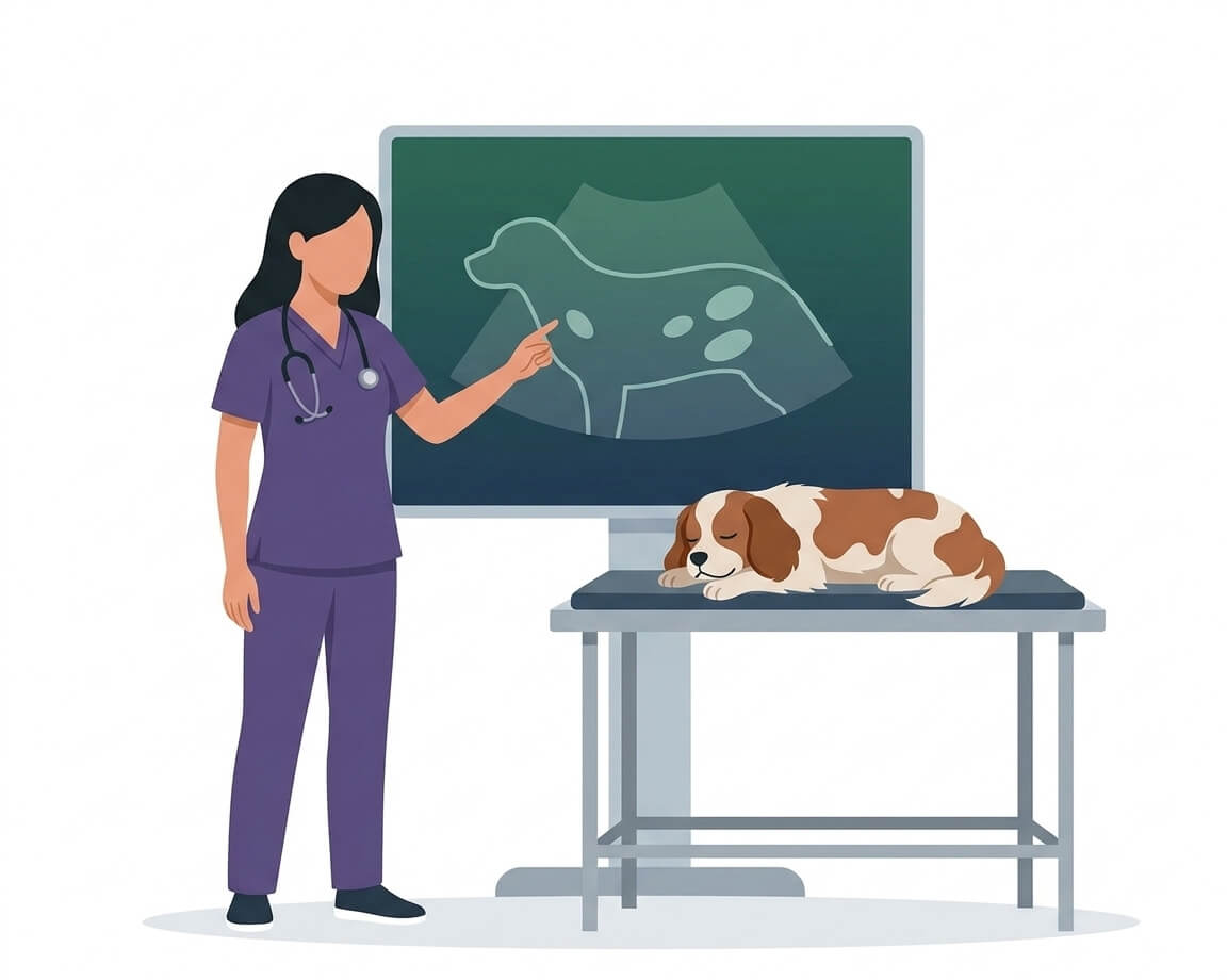
When a vet says “we need to run some imaging tests”, many owners are left with more questions than answers. Will it hurt? Does it need anaesthesia? What exactly are they going to see? It’s a completely understandable reaction — concern for your pet is real, and there isn’t always time during a consultation to explain everything calmly.
This article answers those questions. We explain what a veterinary ultrasound is and what a veterinary X-ray is, what each one is used for, how they differ, and when they’re used together. All in plain language, without unnecessary jargon.
At Rovira Veterinary Clinic we have both veterinary diagnostic imaging techniques available on-site, here in Gràcia, Barcelona. That means there’s no need to refer your pet to an external centre: the tests are carried out here and the results are interpreted during the same visit, before you leave.
Veterinary ultrasound: seeing the organs in real time
What it is and how it works
Ultrasound uses sound waves, not radiation. A transducer — the probe the vet holds against the skin — emits high-frequency sound waves that pass through the tissues and bounce back to the device. The equipment converts those signals into a moving image displayed on a screen in real time.
The procedure is not painful. Your dog or cat will feel nothing more than the gentle pressure of the probe and the coolness of the conductive gel applied to the skin. In the vast majority of cases no sedation is needed: the animal can remain conscious and calm throughout.
One thing worth knowing in advance: the area to be examined needs to be shaved. Fur prevents direct contact between the probe and the skin, which degrades image quality. Many owners are surprised to see their pet clipped, but it’s a necessary and temporary step — the coat grows back with no lasting effects.
What ultrasound is used for in dogs and cats
Ultrasound is the tool of choice when soft tissues need to be examined or abnormal fluid accumulations need to be detected. Unlike an X-ray, it provides information about the texture and internal architecture of organs, not just their outline.
| What is evaluated | When it is indicated |
|---|---|
| Abdominal organs (liver, kidneys, spleen, bladder, uterus) | Persistent vomiting, loss of appetite, abdominal distension, urinary problems |
| Masses or tumours | When something abnormal is felt during physical examination, or unexplained weight loss is present |
| Pregnancy | Confirming a pregnancy and monitoring foetal development |
| Heart (echocardiography) | Heart murmurs detected in consultation, breathing difficulties, excessive fatigue |
| Guided sample collection | Organ cytologies, drainage of fluid accumulations in body cavities |
When investigating digestive, kidney, or reproductive symptoms, ultrasound is typically the first test requested as part of an internal medicine workup, because it provides direct, real-time information about the organs involved.
In the case of the kidneys specifically, ultrasound is one of the key tests for assessing structural changes in cats with suspected chronic kidney disease, one of the most common conditions in cats over seven years of age. If your pet is a cat, we’ve put together a dedicated guide on when a cat needs an ultrasound and what it can detect, covering the kidneys, urinary tract, intestine and feline echocardiography in detail.
Veterinary X-ray: imaging the bones and cavities
What it is and how it works
X-rays use radiation to produce static greyscale images. Denser structures — bones, lungs, metallic foreign bodies — absorb more radiation and appear lighter on the image; soft tissues appear darker. It’s precisely this difference in density that makes X-ray so useful for the skeletal system and the chest.
The procedure is quick and painless. The image is ready within minutes and results are available immediately. In some cases a light sedation may be needed — when the animal is very agitated or the required position is uncomfortable to hold — but this is not the norm.
Radiation exposure during a single diagnostic X-ray is minimal and well within safe limits for the animal. There is no known risk associated with the occasional use that is made in standard veterinary diagnostics.
What X-ray is used for in dogs and cats
X-ray is irreplaceable when it comes to structures that ultrasound cannot visualise well: bones, lungs, and any dense object the animal may have swallowed.
| What is evaluated | When it is indicated |
|---|---|
| Bones and joints | Fractures, luxations, arthritis, hip or elbow dysplasia |
| Lungs and chest | Persistent cough, breathing difficulties, chest trauma |
| Foreign bodies | When ingestion of an object is suspected |
| Spine | Back pain, paralysis, or difficulty moving normally |
| Abdomen | When ultrasound is insufficient or needs to be complemented |
When a pet presents with a sudden limp, a fall, or any suspicion of fracture or bone injury, X-ray is the first test indicated. In the field of veterinary traumatology, it is the essential tool for assessing skeletal integrity and planning treatment.
Ultrasound or X-ray: which one is needed?
The most important answer we can give is this: ultrasound and X-ray are not competing techniques — they complement each other.
Each has its strengths and natural limitations:
- X-ray is superior for dense structures: bones, lungs, metallic foreign bodies, and the overall outline of abdominal organs.
- Ultrasound is superior for soft organs, fluids, moving tissues (like the beating heart), and evaluating the internal texture of the liver, kidneys, or spleen.
- Often, both tests are used in the same visit: the X-ray provides the broad overview and the ultrasound goes deeper into the details. They don’t contradict each other — they complete the picture.
The decision about which test to request — or whether one, both, or neither is needed — is always made by the vet based on the symptoms, clinical history, and physical examination findings. There is no fixed rule: every case is different.
At Rovira we have both diagnostic imaging techniques available on-site in our Gràcia clinic, which means they can be combined in a single appointment when the case calls for it — no waiting, no extra trips.
Frequently asked questions
Will my dog’s fur be shaved for the ultrasound?
In most cases yes, in the specific area to be examined. It’s necessary to achieve good contact between the probe and the skin and obtain a quality image. The fur grows back within a few weeks with no lasting effects.
Does my pet need anaesthesia for an ultrasound or X-ray?
In the vast majority of cases, no. Both procedures are painless and most dogs and cats tolerate them perfectly while fully awake. Only in situations where the animal is very agitated or the positioning is particularly complex might a light sedation be needed.
How long does a veterinary ultrasound or X-ray take?
A standard abdominal ultrasound usually takes between 20 and 40 minutes. An X-ray is faster — the image is ready in a matter of minutes. The total visit time depends on how many areas need to be examined and whether both techniques are combined.
Can I see the results on the same day?
Yes. At Rovira we interpret the tests during the same visit and explain our findings before you leave. There’s no waiting for days or coming back for a result.
Is the X-ray dangerous for my pet?
No. Radiation exposure during a diagnostic X-ray is minimal and completely safe. The level of radiation used in standard veterinary diagnostics carries no known risk with the occasional use involved.
If your pet needs imaging tests or you have questions about what might be going on, the team at Rovira Veterinary Clinic is here to help. You can find more information about our tests on our diagnostic imaging page, or contact us to book an appointment.
Sources
Do you have any questions about your pet?
Book an appointment