
 685 738 325
Diagnostic Imaging
Cat ultrasound: when your cat needs one and what it can detect
24 August 2026 · Updated on 3 September 2026

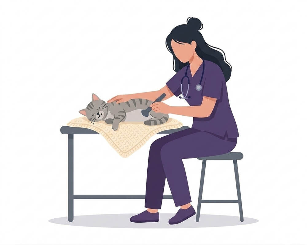
Cats are extraordinarily good at hiding that something is wrong. It’s an inherited instinct: in the wild, showing weakness is an invitation to predators, so an unwell cat tends to hide away, eat a little less and move a little less, without complaining. By the time the change is obvious to us, the problem has usually been there for a while.
That’s exactly the gap an ultrasound fills. It lets us look inside the abdomen and the heart of a cat that appears perfectly fine from the outside, in real time, painlessly and without radiation. Often it confirms that nothing is wrong — and that reassurance counts for something too. Just as often it finds, weeks or months earlier than watching at home ever would, something that can still be managed.
In this guide we explain what a feline ultrasound actually is, how it differs from an X-ray, which signs justify requesting one, what conditions it can detect in each organ, and how to prepare your cat so the day of the scan is as stress-free as possible.
What an ultrasound is and how it differs from an X-ray
An ultrasound works using sound waves: a probe placed against the skin emits high-frequency waves that pass through the tissues and bounce back. The equipment translates those echoes into a moving image the vet watches on screen while scanning. No radiation is involved, there’s no pain, and the cat can stay awake.
The key difference from an X-ray is what kind of information each one gives. An X-ray works with densities: it distinguishes hard from soft very well, which is why it’s irreplaceable for bones, lungs and swallowed objects. But of a kidney it shows you only the outline. An ultrasound, by contrast, goes inside the organ and shows its internal structure: whether the renal cortex has thinned, whether there’s a cyst, whether the liver texture is abnormal, whether an intestinal wall has thickened.
| Ultrasound | X-ray | |
|---|---|---|
| What it uses | Sound waves (no radiation) | X-rays |
| Strong for | Soft organs, fluids, moving tissue | Bones, lungs, dense foreign bodies |
| What it shows of an organ | Its internal structure | Its size and outline |
| Image | Moving, in real time | Static |
| Sedation | Usually not needed | Usually not needed |
When your cat might need an ultrasound
There’s no fixed list, but there is a recurring pattern: an ultrasound comes into play when there’s a persistent symptom with no clear cause, or when blood work comes back abnormal and the question is which organ lies behind that result.
These are the situations that most often lead to one being requested for a cat:
- Weight loss with no change in diet, especially if the cat is still eating normally.
- Chronic vomiting or diarrhoea, or vomiting that recurs every few days over weeks.
- Drinking and urinating more than usual — one of the earliest warnings of kidney trouble in cats.
- Difficulty or pain when urinating, unproductive straining in the litter tray, blood in the urine.
- Loss of appetite lasting more than a couple of days.
- A distended abdomen or one that’s painful on palpation.
- A palpable lump found by the vet during the examination.
- Abnormal blood work: raised urea and creatinine, high liver enzymes, unexplained anaemia.
- A heart murmur, rapid breathing at rest, or a sudden episode of weakness in the hind legs.
- Lethargy, hiding more than usual, or any of the signs that a cat is unwell that persist beyond a few days.
One feline nuance is worth insisting on: in a cat, “he’s just a bit quiet” is a symptom, not an impression. If you’re unsure whether what you’re seeing justifies a visit, the answer is almost always yes.
What an abdominal ultrasound can detect in cats
A complete abdominal study examines, organ by organ, the kidneys, bladder, liver, gallbladder, spleen, pancreas, stomach, intestinal loops, abdominal lymph nodes and adrenal glands. These are the findings that carry most weight in feline practice.
Kidneys: a window onto the most common problem in older cats
The kidney is by far the organ that most often justifies an ultrasound in a cat of a certain age. Chronic kidney disease is one of the most common conditions from seven years onwards, and it has a cruel characteristic: the markers in blood don’t shift until a significant portion of kidney function has already been lost.
An ultrasound provides what blood work cannot — the structure. It shows whether the kidneys have changed size, whether the cortex is thinned or abnormally bright, whether there are cysts, whether there are stones, whether the renal pelvis is dilated because urine isn’t draining properly. And it allows the two kidneys to be compared with each other, which is valuable information in itself.
If your cat already has a kidney diagnosis, or you suspect one, we’d recommend reading our full guide to chronic kidney disease in cats: it covers staging, management and what can be done to slow its progression.
Bladder and lower urinary tract
Lower urinary problems are another feline classic, grouped under the name feline lower urinary tract disease (FLUTD): cats going in and out of the litter tray, straining with no result, urinating outside the tray, or passing blood.
An ultrasound shows whether there are stones, whether crystals have accumulated, whether the bladder wall is thickened by inflammation, whether there are clots, or whether there’s a mass. It also assesses how full the bladder is and rules out an obstruction, which in a male cat is a genuine emergency: if a cat is straining to urinate and producing nothing, this is not something to leave until tomorrow.
A practical detail: the bladder is much easier to see when full, so it helps if your cat hasn’t urinated immediately beforehand.
Liver, spleen and intestine
In the liver, an ultrasound assesses size, texture and outline, and detects nodules, masses, fat accumulation or gallbladder abnormalities. It’s particularly useful in cats that have stopped eating, because a cat with prolonged anorexia can develop secondary liver problems worth identifying early.
In the spleen, it looks for enlargement, nodules and masses.
And in the intestine, feline ultrasound has a very specific role. It measures the thickness of the intestinal wall and evaluates its individual layers. When that wall is thickened, the two most common causes in a cat are inflammatory bowel disease and low-grade intestinal lymphoma — and here it’s worth being honest about the limits of the test: an ultrasound cannot reliably tell one from the other. Certain patterns, such as selective thickening of the muscular layer or enlargement of nearby lymph nodes, tip the suspicion towards lymphoma, but confirmation requires a biopsy.
That doesn’t diminish the value of the ultrasound — quite the opposite. It’s what turns “he vomits a lot and I don’t know why” into “we need to biopsy this specific stretch of intestine”. It points to where to look, and rules out unnecessary tests in other directions.
Pregnancy in unspayed cats
If your cat isn’t spayed and pregnancy is suspected, an ultrasound confirms it at an early stage, allows an estimate of how many foetuses there are, and assesses whether their development and heartbeats are normal. It also detects uterine problems that have nothing to do with pregnancy, such as an accumulation of fluid or pus, which in an older unspayed cat is a possibility that always needs ruling out.
For an exact foetal count, an X-ray in the final stage of pregnancy is more reliable, because the skeletons are visible by then. Another example of the two techniques working together.
Echocardiography: the feline heart and hypertrophic cardiomyopathy
If there’s one chapter where feline ultrasound earns its keep, this is it.
Hypertrophic cardiomyopathy (HCM) is the most common heart condition in cats: it’s estimated to affect around 15% of the general cat population, and that figure rises to close to 29% in geriatric cats. It involves a thickening of the left ventricular muscle that leaves the heart less able to fill with blood.
What makes it such a particular problem is its silence. Many cats with HCM show no visible symptoms whatsoever for years. They don’t cough — cats with heart disease rarely do — they don’t tire noticeably because an indoor cat regulates its own activity, and you can’t even rely on a murmur: there are cats with HCM whose hearts sound entirely normal on auscultation, and cats with an audible murmur whose hearts are perfectly healthy.
Far too often, the first sign of the disease is already a serious complication: pulmonary oedema, or an aortic thromboembolism — a clot that breaks free and blocks the circulation to the hind legs, causing sudden and very painful paralysis.
Echocardiography is the reference test for diagnosing HCM, and the only one that can detect it before a cat shows any symptoms at all. It measures the thickness of the ventricular walls, shows how the chamber fills and empties, assesses the size of the left atrium and detects whether blood is pooling.
Why it matters especially before anaesthesia
A heart with HCM copes less well with the changes in volume and rate that accompany a general anaesthetic. So in a cat with a murmur, with a family history, of a predisposed breed — Maine Coon, Ragdoll, Sphynx, British Shorthair, Persian — or simply of advanced age, knowing the real state of the heart before surgery changes the decisions made in theatre: which drugs are chosen, at what rate fluids are given, what is monitored most closely.
It’s the same logic we set out in our guide to veterinary anaesthesia in dogs and cats: the pre-anaesthetic assessment isn’t a formality, it’s what allows the protocol to be tailored to that particular animal rather than applying a standard one.
How to prepare your cat for an ultrasound
Preparation is straightforward, but doing it properly makes a real difference to image quality.
Fasting beforehand. As a general rule, food is withheld for around 8-12 hours before an abdominal ultrasound. The reason is purely technical: gas and the contents of the stomach and intestine block sound waves and can leave whole areas invisible. Water is normally still allowed. Always confirm the specific instructions for your case with your vet, since guidance differs for diabetic or very young cats.
A full bladder. As mentioned, the bladder is far easier to examine with urine in it. If possible, avoid letting your cat urinate in the hours beforehand.
Shaving. The area being examined is shaved. It’s essential: fur traps air, and without direct contact between the probe and the skin there’s no usable image. Only the necessary area is shaved and the fur grows back with no lasting effects.
Sedation. Not always needed. A calm cat, in a quiet environment and handled unhurriedly, usually tolerates the scan well while awake. Sedation is considered when the cat is very stressed or in pain, when the required position is difficult to hold, or when a sample has to be taken under ultrasound guidance. The decision is made case by case.
The journey and arrival. This is where you can help most. Leave the carrier open at home for a few days beforehand so it stops being “the going-to-the-vet box”. Cover the carrier with a towel during the journey: fewer visual stimuli, less stress. And put a blanket that smells of home inside.
Duration. The scan itself usually takes between 20 and 60 minutes, depending on how many organs need reviewing and how cooperative the patient is.
Frequently asked questions
Does an ultrasound hurt my cat?
No. It’s a painless procedure: no needles, no incisions and no radiation. Your cat only feels the gentle pressure of the probe and the coolness of the conductive gel. What can be uncomfortable isn’t the scan but lying still and being held for a while somewhere unfamiliar — and that’s managed with patience and low-stress handling.
Does my cat need sedation for an ultrasound?
In many cases, no. A calm cat tolerates an ultrasound perfectly well while awake. Sedation is considered when the cat is very stressed or in pain, when a specific position has to be held for a long time, or when a sample needs taking under ultrasound guidance. It’s an individual decision.
How long does a cat ultrasound take?
Between 20 and 60 minutes, depending on what’s examined. A full abdominal study sits at the upper end; a scan aimed at one specific question is quicker. Preparation and shaving add to that.
How often should my senior cat have check-ups?
From around seven years of age, the recommendation shifts from an annual check-up to one every six months. An ultrasound isn’t part of every one of those visits: it’s added when the examination or blood work shows something worth looking at from the inside. You can see the intervals by life stage in our guide to how often to take your pet to the vet.
Does my cat need to be shaved?
Yes, in the area being examined. Fur prevents contact between the probe and the skin, and without that contact there’s no quality image. Only the necessary area is shaved, not the whole cat, and it grows back within a few weeks.
Can an ultrasound rule out a tumour?
It can detect and locate one, but it can’t name it. It shows that a mass is there, where it is and how big it is. Determining what type it is requires a fine needle aspirate guided by ultrasound, or a biopsy.
Can an ultrasound replace a blood test?
No, they’re complementary. Blood work tells you how the organs are functioning; an ultrasound tells you what they look like inside. A cat can have structurally abnormal kidneys with blood work that’s still normal, and the other way round. The two are normally interpreted together.
Does my indoor cat need one too?
Living indoors protects against accidents, fights and many infections, but not against kidney disease, HCM or intestinal lymphoma. In fact, in an indoor cat changes in activity are harder to spot, precisely because their routine is quieter to begin with.
If you have doubts about your cat, don’t wait for it to become obvious
With cats, waiting until you’re certain something is wrong usually means waiting too long. An ultrasound is a quick, painless, radiation-free test that often settles the question during the same visit — either because it finds something in time, or because it confirms there’s nothing to worry about.
At Rovira Veterinary Clinic we have diagnostic imaging available on-site, here in Gràcia, Barcelona, and we approach feline handling with low-stress techniques as part of our feline medicine service.
If you’ve noticed something odd about your cat, or you simply want to bring their check-ups up to date, get in touch and we’ll take a calm look together.
Sources
Do you have any questions about your pet?
Book an appointment