
 685 738 325
Diagnostic Imaging
Cat X-rays: when your cat needs one and what it reveals
8 September 2026 · Updated on 8 September 2026
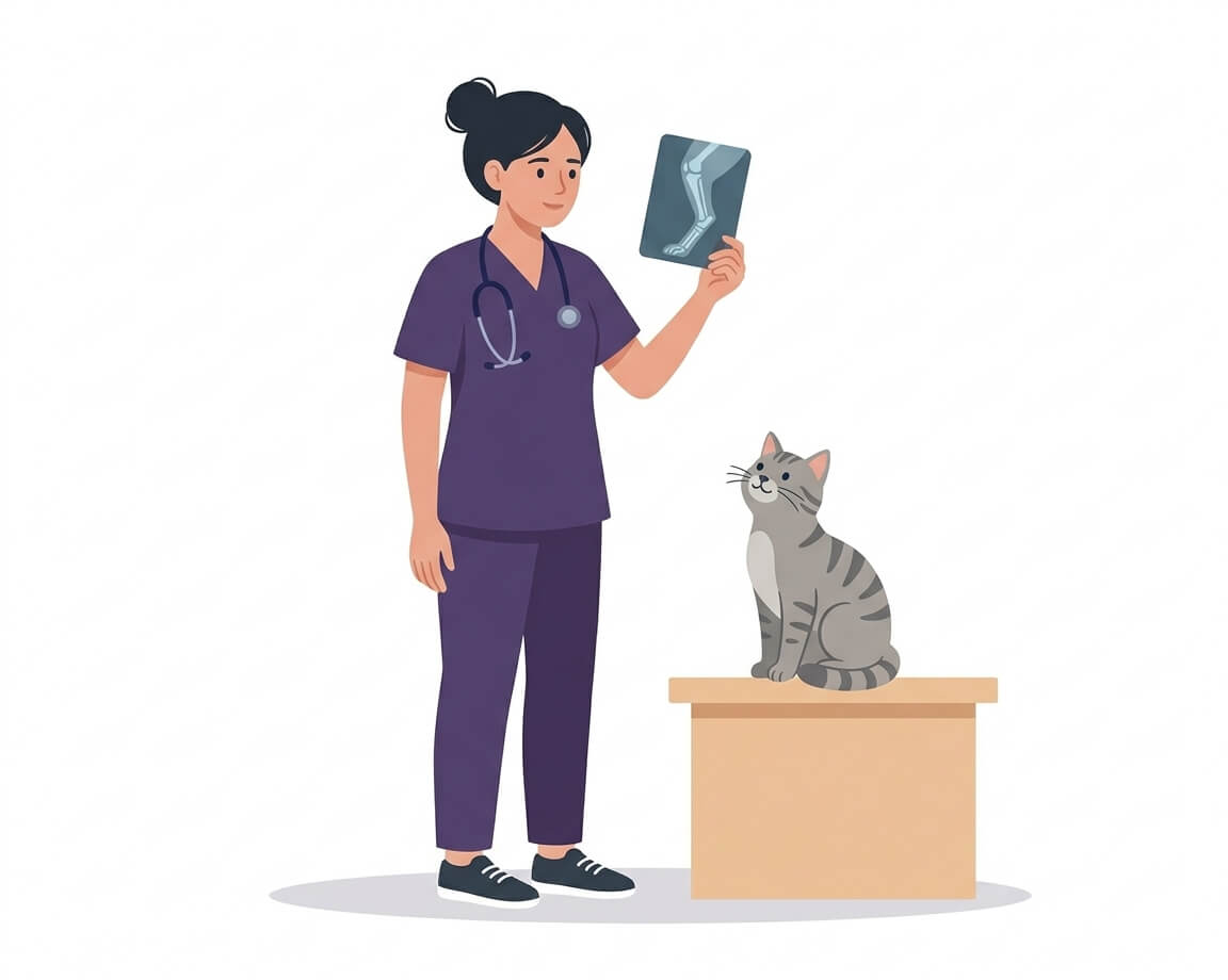
Cats are said to have nine lives. It’s a nice line, and there’s something to it: a cat is an extraordinarily resilient animal, able to fall from a considerable height and get up as if nothing had happened, or to live for months with a pain that any other animal would have announced at the top of its voice. The trouble is that this same resilience is exactly what throws us off.
Because there are very specific situations in which a cat that “seems fine” is not. A cat who has fallen off the balcony and walks normally may have a bruised lung. A cat who has been playing with a hair tie and is only vomiting a little may have an intestine folding in on itself. A cat who eats without complaint may have teeth dissolving from the inside. In all of those cases, the difference between a scare and a genuine emergency comes down to an image.
This guide explains when a cat X-ray is the right test — rather than an ultrasound — what exactly it reveals in each situation, and which signs at home should have you reaching for the carrier without waiting until tomorrow.
Cat X-ray vs ultrasound: when each one is used
It’s the question that comes up most often in the consulting room, and the answer is simpler than it looks: each test sees well what the other sees badly.
An X-ray works with densities. X-rays pass through tissue, and the denser ones — bone, metal — slow the beam more and come out white in the image; air lets it through almost entirely and comes out black. That makes it the irreplaceable test for three things: bone, air and radiopaque objects.
An ultrasound, by contrast, works with sound waves that reflect off the interfaces between tissues. It can go inside a soft organ and show its internal architecture in motion. But it stops dead at bone and at air — which is why it is no use for assessing an air-filled lung or for looking inside a joint.
| X-ray | Ultrasound | |
|---|---|---|
| Sees very well | Bone, lungs, chest, dense objects, stones | Inside of soft organs, fluid |
| Sees poorly or not at all | Internal structure of soft organs | Bone, air-filled lung, intestinal gas |
| Type of image | Static, panoramic | Moving, real time |
| Radiation | Yes, at a very low dose | No |
| Typical use in cats | Trauma, chest, foreign bodies, teeth, stones | Kidneys, liver, intestine, bladder, heart |
| Approximate duration | 10-20 minutes | 20-60 minutes |
Foreign bodies: the most common reason for an emergency
Cats don’t usually swallow a whole ball the way a dog does. Their thing is linear objects: a length of sewing thread, a hair tie, gift ribbon, a hoodie drawstring, tinsel from the Christmas tree. And that, far from being less serious, is considerably more dangerous.
Why thread is more dangerous than a solid object
The anatomy of the cat’s tongue is largely to blame. It is covered in backward-facing papillae, like tiny hooks, which make it physically very difficult to spit something out once it has entered the mouth: the only thing the cat can do is keep swallowing.
When what they swallow is thread, something specific happens: one end usually becomes anchored, very often caught around the base of the tongue. The other end carries on down the digestive tract, dragged along by the normal movement of the intestine. Because the thread can’t advance fully, the intestine gathers and folds in on itself around it, like a curtain bunching on its rail. This is known as intestinal plication.
And the taut thread starts to behave like a saw. Over the following hours it can cut through the intestinal wall from the inside and perforate it, spilling intestinal contents into the abdomen — peritonitis. This is why a linear foreign body in a cat is not an ordinary obstruction: it is a genuine surgical emergency, and time matters.
What the X-ray sees, and what it doesn’t
Here it’s worth being honest, because this is where a lot of articles oversimplify:
- Radiopaque objects — metal, needles, pins, some bones, certain dense plastics, stones — show up directly on the X-ray, with their exact shape and position. A swallowed safety pin appears drawn on the image.
- Radiolucent objects — thread, fabric, rubber, soft plastic — do not show up. The X-ray doesn’t show the thread: it shows what the thread is doing to the intestine. The radiologist looks for indirect signs: bunched or pleated loops of bowel, gas bubbles with odd shapes (elongated, comma-shaped, eccentric within the loop) and an overall pattern that doesn’t fit a normal intestine.
When those signs are subtle, ultrasound is usually the next step, because in trained hands it can directly show the plication and even the material itself inside the intestinal lumen. Another example of the two tests working together rather than competing.
Signs at home that should prompt a call
- Repeated vomiting, especially if water comes back up too.
- A sudden refusal to eat, in a cat who was eating normally.
- Drooling or odd movements of the mouth, dry swallowing.
- A hunched posture, or objecting to having their belly touched.
- No longer passing stools, or straining without result in the litter tray.
- Marked apathy and hiding — often the first warning, and the easiest to miss.
One thing you can try at home before you leave: gently open their mouth and look under the tongue, which is where the thread tends to anchor. If you can see it, saying so the moment you arrive at the clinic speeds things up considerably.
But only do this if your cat lets you: if they resist, leave it and head straight to the clinic. The base of the tongue is a difficult area to see even in the consulting room — it often takes a tongue depressor, a light and, in many cats, light sedation — so wrestling with it at home has little to gain and quite a lot to lose: you may get bitten, and more importantly you may over-stress a cat whose abdomen or breathing may already be compromised. Seeing nothing doesn’t rule anything out either.
High-rise syndrome: the “parachuting cat”
This is the name given to the set of injuries a cat sustains when falling from a window, a balcony or a terrace. It’s a classic of urban feline practice, and in vertical cities like Barcelona it turns up as an emergency every summer, when the windows go open.
It’s almost never carelessness on the cat’s part: the usual sequence is that they were hunting — a bird, a fly, a moth — or that they dozed off on a narrow railing and slipped while stretching. A cat focused on prey stops calculating the edge.
What happens to a cat’s body during a fall
Cats have a remarkable righting reflex: in mid-air they can rotate their body using the spine and tail until their feet point downwards, and they reach impact with the limbs flexed, absorbing some of the energy. It’s the reason they survive falls that would be fatal to other animals of their size — in the published clinical series, the majority of cats who reach the vet after a fall survive.
But surviving is not the same as walking away unharmed. The injuries that recur most often are:
- Limb fractures, more common in the hind legs than the front.
- Jaw fractures and separation of the hard palate from the chin striking the ground — very characteristic of this syndrome.
- Thoracic trauma: pneumothorax (free air in the chest, collapsing the lung) and pulmonary contusion (the lung bruised and bleeding internally). This is the injury that drives the prognosis, and it affects a very substantial share of the cats seen.
- Ruptured bladder, particularly if the cat fell with a full bladder.
- Head trauma, internal bleeding and diaphragmatic tears.
The counter-intuitive point about height
There’s a widely repeated idea — based on a classic study from the 1980s — that falls from lower floors can be more injurious than falls from higher ones. The proposed explanation is that the cat needs a certain amount of falling time to complete the rotation and then to relax and spread the body out, increasing air resistance like a parachute. In a short fall there isn’t time: they hit the ground mid-turn and with a rigid body.
It’s worth adding the caveat that almost nobody mentions: this finding is disputed. Later studies have found the opposite — more severe injuries with greater height — and an obvious bias in the original data has been pointed out, since cats who die in a fall from a very high floor often never reach the clinic at all and never enter the statistics.
For you as an owner the scientific debate is beside the point, because the practical conclusion is the same either way: there is no height from which falling is safe. A second floor doesn’t rule out a serious injury, and a seventh doesn’t guarantee one.
What to do right after a fall
- Move them as little as possible. If you suspect a fracture or a spinal injury, slide them onto a rigid surface — a board, a baking tray, the base of a dismantled carrier — rather than picking them up in your arms.
- Don’t give food or water. If sedation or surgery is needed, an empty stomach works in their favour.
- Don’t give any human painkiller. Paracetamol is lethal to cats, and ibuprofen is not an option either.
- Call the clinic to say you’re on your way, so they’re ready for you.
- Take them even if they seem fine. Yes, again: it’s the item on this list that saves the most lives.
Prevention: screens, not railings
The only prevention that genuinely works is physical. A railing is not a barrier to a cat, and a window left “just a bit open” on a tilt-and-turn setting is particularly dangerous: the cat slips through the gap at the top, slides down and becomes trapped by the abdomen in the wedge, a situation that causes severe spinal and internal organ injuries even without a fall.
What works are sturdy insect screens or protective mesh on windows and balconies, and dedicated guards for tilt-and-turn windows. It’s a small investment that solves the whole problem, and it matters especially if yours is an indoor cat with no experience of real heights.
Chest problems: lungs and heart
If there’s one territory where the X-ray has no rival, it’s the chest. The lung is full of air, and air is precisely the ultrasound’s blind spot — which is why the chest X-ray remains the first-line test for any cat with breathing difficulty.
A chest X-ray assesses:
- The lung pattern: whether there is fluid within the lung (oedema), inflamed or infected areas, blood from a contusion, or masses.
- The pleural space: whether air has accumulated around the lung (pneumothorax) or fluid is compressing it (pleural effusion), two situations that suffocate the cat from outside the lung.
- The cardiac silhouette: the size and shape of the heart, and the appearance of the great vessels.
- The diaphragm, to rule out a diaphragmatic hernia after trauma.
- The ribs and thoracic spine.
When to suspect a breathing problem in a cat
Cats hide respiratory distress to a degree that is genuinely surprising. These are the signs that should not wait until tomorrow:
- Abdominal breathing: the belly moves markedly with each breath, with visible effort.
- Open-mouth breathing at rest. In a dog it’s normal; in a cat it is an emergency, almost always.
- A high resting or sleeping respiratory rate. It’s easy to measure at home: count the chest movements over 30 seconds and double it. With the cat calm or asleep, a sustained rate above 30 breaths per minute warrants a call.
- A sphinx posture with the neck extended and the elbows held away from the body, refusing to lie on their side.
- Pale or bluish gums or tongue.
X-rays and the heart: how far they go
A chest X-ray shows whether the heart is enlarged and, above all, whether the lung has become waterlogged as a result. That makes it indispensable in an emergency. But it has an important limitation in cats: the most common feline heart disease, hypertrophic cardiomyopathy, thickens the muscle inwards into the chamber, so in the early stages a cat can have a diseased heart and a completely normal-looking cardiac silhouette on the X-ray.
In advanced stages that changes, and the X-ray does offer clues: when the left atrium dilates, the cardiac silhouette seen from above takes on a characteristic shape vets call the “valentine heart”. The problem is that by then the disease has been established for some time — and the whole point was to catch it before that.
That’s why, for a cat’s heart, the reference test is the echocardiogram, which does measure wall thickness and chamber size. We explain it in detail in our cat ultrasound guide. In practice, a cat who is breathing badly usually gets both: the X-ray says what is happening to the lung, and the echocardiogram says why.
Urinary obstruction in male cats
A male cat straining in the litter tray without producing urine is a life-threatening emergency, not a behavioural problem or constipation. The male urethra is long and narrow, and when it becomes blocked — by crystals, a plug of mucus and cells, or a stone — urine stops coming out. Within hours, toxins and potassium build up in the blood, with a real risk of acute kidney failure and cardiac arrest. The margin is measured in hours, not days.
The warning signs:
- Going in and out of the litter tray repeatedly with no result.
- Straining and producing nothing, or just a few drops, sometimes with blood.
- Crying or vocalising while trying.
- Persistent licking of the genital area.
- Vomiting, hiding, refusing food, lying around listlessly.
Here the X-ray contributes something very specific. The two most common stones in cats are calcium oxalate and struvite, and both usually show up on the image: oxalate stones are clearly radiopaque, and struvite ones generally are too, although very small or mixed-composition stones can sit at the edge of visibility. Above all, the X-ray covers the entire tract, from the kidneys to the urethra including its pelvic course — precisely where blockages most often lodge, and where ultrasound has great difficulty reaching. There, the X-ray is unbeatable.
Ultrasound plays the complementary role: inside the bladder it detects small uroliths better, whatever their mineral composition, and it also assesses wall thickness, contents and the state of the kidneys. Once again, the two together.
Bones, joints and dental X-rays
The musculoskeletal system
For bone there is no alternative: fractures, dislocations, arthritis, dysplasia and spinal injuries are diagnosed with X-rays. Ultrasound stops at the bone surface and cannot go in.
In cats, moreover, lameness is hard to spot. A cat with joint pain rarely limps in any obvious way: what they do is stop jumping, choose lower places to sleep, get onto the sofa in two stages instead of one leap, use the scratching post less, groom themselves less thoroughly, or start missing the litter tray because climbing in has become difficult. Feline osteoarthritis is very common in older cats and hugely underdiagnosed, precisely because these changes get put down to “just getting old”. They are reason enough for an X-ray.
Dental X-rays: the big blind spot
If there’s one use of radiography that changes a cat’s life and that almost nobody knows about, it’s this one.
Feline tooth resorption is one of the most common oral diseases in cats: depending on the series consulted, it affects somewhere between 20% and 60% of the feline population, and the proportion rises sharply from around five or six years of age. The tooth’s own tissue is progressively destroyed from within, leaving the dentine exposed. It is intensely painful.
And here’s the problem: a good proportion of the lesions cannot be seen by looking in the mouth. They start below the gum line or inside the root, where no visual examination can reach. Studies comparing visual examination with dental radiography find that X-rays detect a far greater number of affected teeth — in cats with no visible lesion at all, radiographs reveal lesions in a high proportion of cases.
The cat, meanwhile, carries on eating. Not going off their food does not mean not being in pain: it means eating takes priority. The real signs tend to be subtler: chewing on one side only, dropping food, chattering the jaw while eating, drooling, marked bad breath, grooming less, or becoming grumpy when their head is touched.
That’s why, in a properly performed dental procedure in a cat, full-mouth dental radiographs are not an optional extra: they are what makes it possible to decide which tooth can be saved and which has to come out, and to avoid leaving behind a root that would carry on hurting. Our veterinary dentistry service covers dental cleaning and tooth extractions under anaesthesia; we do not have intraoral dental radiography, so a case that needs it is best assessed at a centre that does.
Does my cat need sedation for an X-ray?
In most cases, no. An X-ray is quick and painless: no needles, no incisions, and with digital equipment the image appears on screen in seconds. A cooperative cat is placed on the table, held gently for just as long as it takes, and the exposure is made.
Sedation is considered in specific situations, always assessing the individual case:
- When there is pain: moving a fractured leg into the right position hurts, and sedating is the humane thing to do.
- When the positioning is demanding: some orthopaedic and spinal views require a very precise alignment that an awake cat will not hold.
- When the cat is very stressed or fear-aggressive, to the point where pressing on would be worse for them than sedation.
- For dental X-rays, which are always done under general anaesthesia because the sensor has to be placed inside the mouth; this normally uses the same anaesthetic as the dental cleaning.
- When movement ruins the image and the study would have to be repeated several times, adding up exposures and stress.
Which drugs are used, at what doses and with what monitoring is strictly a veterinary decision, made after assessing each cat’s age, kidney status, heart and the reason for the study. If you’d like to understand how a sedation or anaesthetic is prepared and monitored, we go into it in our guide to veterinary anaesthesia in dogs and cats.
And there is a lot you can do to make it unnecessary: half of a cat’s stress at the clinic is generated before they arrive. Leaving the carrier open at home as just another piece of furniture, covering it with a towel for the journey, putting a blanket that smells of them inside, and using synthetic facial pheromones 15-20 minutes beforehand make an enormous difference. We set it out step by step in how to take your cat to the vet without stress.
Frequently asked questions
Is the radiation from an X-ray dangerous for my cat?
No. The X-ray dose from a diagnostic radiograph is very low, far below the levels capable of causing harm, and today’s digital equipment needs less radiation still than the old film systems. Standard protective measures are always in place: the beam is limited to the area being studied and staff wear shielding. For the occasional diagnostic use made of it in veterinary practice, the risk is negligible against the benefit of knowing what is happening to your cat.
What should I do if my cat has fallen from a window and seems fine?
Take them to the vet the same day, even if they’re walking and look normal. A pneumothorax, a lung contusion, a ruptured bladder or a fractured palate may not declare themselves for hours. In the meantime: move them as little as possible, don’t give food or water, don’t give any human painkiller, and call the clinic to say you’re on your way. The minimum recommended check is a chest X-ray.
How do I know if my cat has swallowed something?
Repeated vomiting — especially if water comes back up too — a sudden refusal to eat, drooling, a hunched posture, abdominal pain and no longer passing stools. If you suspect thread, a hair tie or ribbon, you can try to look under the tongue, where linear foreign bodies tend to anchor in cats — but only if they let you: if they resist, don’t force it and go straight to the emergency clinic. Seeing nothing doesn’t rule anything out, and the clinic can examine that area with better tools. And if you see thread, don’t pull on it.
How long does the study take?
Taking the image is a matter of seconds and with digital equipment it appears on screen instantly. Allowing for positioning, the various views needed and interpretation, a radiographic study is usually done in 10-20 minutes, within the same visit.
Does my cat need to fast before an X-ray?
For a plain X-ray, no: unlike an abdominal ultrasound, gas and digestive contents don’t prevent a usable image. Fasting is requested when sedation or anaesthesia is anticipated, or if the study will involve contrast. Worth confirming when you book.
Can an X-ray detect a tumour in my cat?
It can detect chest masses, bone masses and abnormal abdominal silhouettes, and it is the reference test for looking for lung metastases before oncological surgery. What it cannot do is say what type of tumour it is: that requires a fine-needle aspirate or a biopsy. And for a mass in a soft abdominal organ, ultrasound defines it far better.
Is an X-ray useful for looking at my cat’s kidneys or liver?
Only partly. It shows size, position and outline, and it detects radiopaque stones, but it doesn’t show the organ’s internal structure. To assess the inside of a kidney, a liver or an intestinal wall, the right test is an ultrasound.
How often should my cat have an X-ray?
It isn’t a routine test: it’s done when there’s a specific reason — trauma, a respiratory sign, a suspected foreign body, lameness, a dental procedure. What is worth keeping up is the regular check-up: annually in adult cats and every six months from around seven years of age, which is when the findings that lead to requesting the test start to appear. You can see the intervals by life stage in our guide to how often to take your pet to the vet.
With a cat, when in doubt, the answer is almost always yes
Cats don’t exaggerate their symptoms: they hide them. Which is why, with them, the “let’s see if they’re better tomorrow” approach fails more often than it works. A fall, repeated vomiting, faster-than-normal breathing or a jump they’ve stopped making are all reason enough for a check — and often a single image settles the question within the same visit.
At Clínica Veterinaria Rovira we have digital radiography and ultrasound on site, in Gràcia (Barcelona). That means we don’t refer your cat to an outside centre: the test is done here and the result is interpreted before you go home. You can see everything our diagnostic imaging service covers, along with our dedicated feline medicine work, where we apply low-stress handling to every procedure.
If your cat has fallen, swallowed something, is breathing oddly or has simply “not been themselves” for a while, get in touch and we’ll take a calm look.
Sources
Do you have any questions about your pet?
Book an appointment